
Neeka Research Library
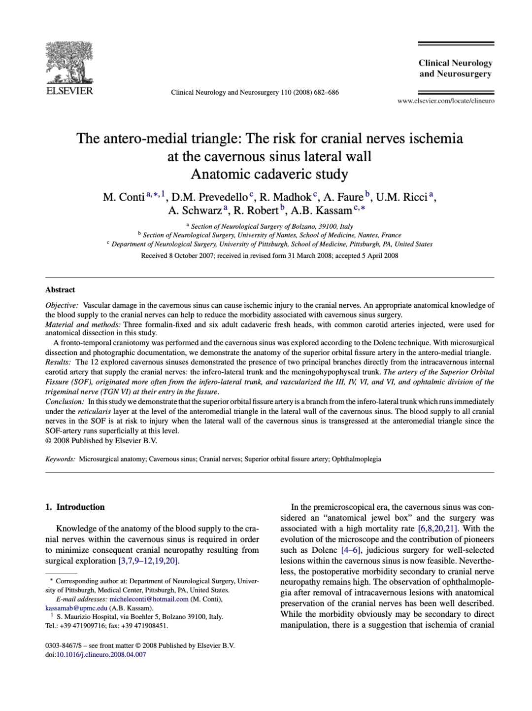
The antero-medial triangle: The risk for cranial nerves ischemia at the cavernous sinus lateral wall


Minimally Invasive Endoscopic Pericranial Flap: A New Method for Endonasal Skull Base Reconstruction


The Posterior Pedicle Inferior Turbinate Flap: A New Vascularized Flap for Skull Base Reconstruction

Address
135 Great George St
Charlottetown, PEI
C1A 4K7
info@neekahealth.com